Congenital ptosis
Ptosis can affect one eye or both eyes. Ptosis may be present at birth, or may be acquired later in life. If a droopy eyelid is present at birth or within the first year of life, the condition is called congenital ptosis. In most cases of congenital ptosis, the problem is isolated and does not affect the vision. Any ptosis that develops over a period of days or weeks can signal a serious medical problem and needs further neurologic and physical evaluation.Congenital Ptosis (Droopy Eyelids in Infants, Toddlers, Children) Etiology (Causes):
In most cases of congenital ptosis, the cause is idiopathic (unknown).
The eyelids are elevated by the contraction of the levator palpebrae superioris. In most cases of congenital ptosis, a droopy eyelid results from a localized myogenic dysgenesis. Rather than normal muscle fibers, fibrous and adipose tissues are present in the muscle belly, diminishing the ability of the levator to contract and relax. Therefore, the condition is commonly called congenital myogenic ptosis (Droopy eyelids causes by a defective eyelid muscle).
Congenital ptosis can also occur when the innervation to the levator is interrupted through neurologic or neuromuscular junction dysfunction.
Histologically, the levator muscles of patients with congenital ptosis are dystrophic (developmentally, they are not normal). The levator muscle and aponeurosis tissues appear to be infiltrated or replaced by fat and fibrous tissue. In severe cases, little or no striated muscle can be identified at the time of surgery. This suggests that congenital ptosis is secondary to local developmental defects in muscle structure.
Congenital ptosis may occur through autosomal dominant inheritance. Common familial occurrences suggest that genetic or chromosomal defects are likely – Genetic causes are likely for their occurence.
Other potential causes of congenital blepharoptosis include:
- Blepharophimosis syndrome: This condition consists of short palpebral fissures (eyelid openings), congenital ptosis, epicanthus inversus, and telecanthus.
•Third cranial nerve palsy (paralysis): Signs of aberrant regeneration are usually present, leading to abnormal movements of the eyelid. The pupil may be paradoxically small and nonreactive.
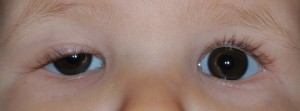
•Horner syndrome: Ipsilateral findings of mild ptosis, miosis, and anhidrosis characterize this syndrome. The ipsilateral lower eyelid may be elevated. Also, because of the lack of sympathetic innervation to the iris melanocyte development, a difference in the iris color between the two eyes may result (called heterochromia).
•Marcus Gunn jaw-winking syndrome: The motor nerve to the external pterygoid muscle is misdirected to the ipsilateral levator muscle. Lid elevation occurs with mastication (chewing) or with movement of the jaw to the opposite side.
•Birth trauma
•Duane syndrome: In this condition, the sixth cranial nerve fails to innervate a lateral rectus muscle. Then, the muscle acquires an innervation of the third cranial nerve. Although the synkinesis produced does not involve lid innervation, enophthalmos with apparent ptosis may result. In Esotropic Duane syndrome, the upper eyelid droops further and the lower lid elevates when the eye is adducted because of a co-contraction of the horizontal rectus muscles.
•Periorbital tumor: Tumors in the orbit (bony socket around the eye) like Neuroblastoma, plexiform neuromas,lymphomas, leukemias,rhabdomyosarcomas, neuromas, neurofibromas, or other deep orbital tumors may produce ptosis or proptosis.
•Kearns-Sayre syndrome: This mitochondrial deletion disorder is characterized by progressive external ophthalmoplegia, heart block, retinitis pigmentosa, and central nervous system manifestations. This condition begins in childhood but is rarely present at birth. The conditions are most likely to become symptomatic in the first or second decade of life. Bilateral ptosis is a prominent feature of this syndrome.
•Myotonic dystrophy: Patients with this condition may present with polychromatic cataracts, gonadal atrophy, or premature thinning and/or loss of hair. Myotonic dystrophy is an autosomal dominant disorder that is characterized clinically by myotonia and progressive muscular weakness.
•Blepharochalasis: This condition is characterized by infiltrative processes that thicken the lids and produce ptosis.
•Myasthenia gravis: A defect at the neuromuscular junction produces relative unresponsiveness to released acetylcholine, resulting in ptosis.
•Pseudotumor of the orbit: Patients with this condition may present with ptosis due to inflammation and edema of the eyelid.
•Pseudoptosis: Less tissue in the orbit (eg, unilateral smaller eye, fat atrophy, blowout fracture) produces the appearance of ptosis secondary to the decreased volume of orbital contents.
Congenital Ptosis (Drooping Eyelids in Infants, Toddlers, Children) Complications:
Amblyopia (lazy eye, in which vision does not develop normally) may result from obscuration of the vision directly or from development of astigmatism indirectly. Development of amblyopia is an indication for immediate surgical correction of the blepharoptosis.
- Occlusion amblyopia (Obstruction of light entering the eye causes a lazy eye and abnormal visual development)
•Astigmatism (visual refractive errors, requiring glasses for correction) from the compression of the droopy eyelid
•Ocular torticollis (Torsion of the neck, as the child turns the neck to see clearly)
Congenital Ptosis